How Technology Is Changing Periodontal Treatment Today

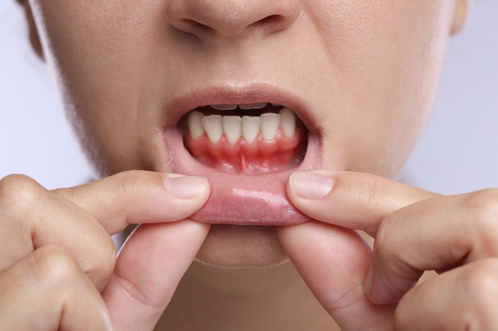

Periodontal treatment used to feel, from the patient’s side, like a narrow set of options. A person came in with bleeding gums, loose teeth, bad breath, or recession. The clinician probed around each tooth, took a few X-rays, diagnosed gum disease, and moved into a familiar sequence of deep cleaning, surgery if needed, and maintenance. That basic framework still exists because the biology of periodontal disease has not changed. Plaque still triggers inflammation. Bone still resorbs when disease progresses. Tissue still heals according to the limits of blood supply, oral hygiene, and systemic health.
What has changed is how precisely we can see disease, how conservatively we can treat it, and how closely we can track healing afterward. Technology has not replaced sound diagnosis or careful hands. It has, however, sharpened both. In the best practices, modern tools are making periodontal treatment more accurate, more comfortable, and in many cases more predictable than it was a generation ago.
That progress matters because periodontal disease is often quiet until it becomes serious. Many patients are surprised to learn they have deep pockets or significant bone loss because they are not in pain. By the time symptoms become obvious, the condition may already involve complex inflammation, tooth mobility, furcation defects, or bite changes. Better tools help clinicians catch problems earlier and intervene with less damage already done.
Seeing more than probing depths
The periodontal probe remains essential. It is simple, inexpensive, and clinically useful. Yet anyone who has practiced for a while knows its limits. Probing depths vary with inflammation, tissue tone, insertion pressure, and patient comfort. Bleeding on probing is meaningful, but it does not tell the whole story. Traditional two-dimensional radiographs can miss important architecture, especially when defects are early or hidden by overlapping structures.
Digital imaging has changed that landscape. Standard digital dental radiographs offer immediate viewing, better image manipulation, and easier comparison over time. For periodontal treatment, that means subtle crestal bone changes are easier to monitor, and patient education improves because people can actually see what the clinician is describing. That sounds small, but it often shifts compliance. A patient who sees vertical bone loss next to a molar usually understands the urgency far better than one who hears a verbal explanation alone.
Cone beam computed tomography, or CBCT, has added another layer. It is not needed for every periodontal case, and thoughtful clinicians avoid using it indiscriminately. Radiation exposure, cost, and case relevance still matter. But when used well, CBCT can reveal the exact shape of intrabony defects, furcation involvement, root proximity, dehiscence, fenestration, and the relationship between periodontal destruction and adjacent anatomy. It is especially helpful when treatment planning regenerative procedures, evaluating compromised molars, or distinguishing endodontic-periodontal problems from isolated periodontal lesions.
A practical example makes the point clearer. A lower molar may look salvageable on a standard radiograph, with what appears to be generalized horizontal loss. On CBCT, the same tooth may show a narrow three-wall defect on one surface, making regeneration plausible, or severe through-and-through furcation destruction, making long-term prognosis much worse. That difference changes the conversation completely. Technology in this setting does not merely decorate the diagnosis. It alters the treatment decision.
Digital charting and better disease tracking
One of the quiet revolutions in periodontal care has been digital charting. This is less glamorous than lasers or three-dimensional imaging, but in day-to-day practice it may have just as much impact. Digital periodontal charting systems can record pocket depths, recession, mobility, furcation involvement, bleeding, and plaque scores more efficiently than handwritten charts. More importantly, they make change over time easier to recognize.
Periodontal disease management is not a one-visit event. It is longitudinal. A six-millimeter pocket that becomes four millimeters with no bleeding after therapy is a clinical success, even if it is not a perfect site. A three-millimeter site that bleeds repeatedly over several recalls may be a warning sign despite looking mild on paper. Technology helps organize these patterns, and pattern recognition is central to good periodontal treatment.
Many practices now overlay charting data with radiographs, photographs, and treatment notes in a way that creates a much richer clinical story. This becomes particularly useful for medically complex patients, smokers, patients with diabetes, or people with a long history of inconsistent home care. Their disease activity rarely follows a simple script. Trends matter more than snapshots.
Intraoral cameras have changed patient acceptance
If there is one technology that routinely changes behavior, it is the intraoral camera. Patients often understand their gums only after they see them close up. Inflamed papillae, plaque buildup near the gingival margin, recession grooves, exposed root surfaces, and bleeding points become concrete rather than abstract.
This matters because periodontal treatment depends heavily on patient participation. Root planing can disrupt biofilm below the gumline. Surgery can reduce pockets or regenerate tissue in selected cases. Maintenance visits can control recurrence. None of that fully succeeds if plaque control at home remains poor. Intraoral photographs create a visual baseline and make progress measurable. When a patient sees less redness and firmer tissue after improved brushing, interdental cleaning, or smoking reduction, the benefit feels real.
Experienced clinicians also use these images to show nuance. A patient may believe aggressive https://devinfthu410.summitviewdaily.com/posts/how-periodontal-treatment-protects-against-future-tooth-loss brushing is helping because the teeth feel clean, while photos reveal abrasion and progressing recession. Another may think “no pain” means “no problem,” while images show clear inflammation. Better communication is not an accessory to treatment. It is treatment.
Ultrasonics and the move toward less traumatic debridement
Scaling and root planing remain foundational in nonsurgical periodontal treatment. What has changed is how often clinicians can perform this therapy more efficiently and with less tissue trauma. Ultrasonic instrumentation, when used skillfully, removes deposits and disrupts subgingival biofilm in a way that can reduce operator fatigue and improve access in deeper pockets.
The technology itself is not new, but modern ultrasonic systems are more refined than earlier generations. Slim tips, better irrigation, adjustable power settings, and improved ergonomics allow for gentler treatment around roots, implants, and inflamed tissues. For patients, this often means shorter appointments, less scraping sensation, and in some cases a smoother postoperative recovery.
There are trade-offs. Heavy tenacious calculus may still require hand instrumentation for complete root surface management. Sensitive patients may respond differently to ultrasonic vibration or water lavage. Clinicians also need excellent tactile discipline because it is easy to overestimate what has been removed if they rely too much on the device and not enough on direct assessment. Technology can improve periodontal treatment, but only when paired with sound technique.
Lasers in periodontal care, useful but not universal
Few technologies in dentistry generate more mixed expectations than lasers. Some marketing around them has outrun the evidence, which is why careful discussion is necessary. Lasers can be helpful in selected aspects of periodontal treatment. They may aid soft tissue decontamination, sulcular debridement, bacterial reduction, and certain surgical procedures. Some patients appreciate the reduced bleeding and more comfortable soft tissue management associated with specific laser applications.
Still, lasers are not a magic substitute for diagnosis, mechanical debridement, or periodontal surgery where surgery is clearly indicated. If a patient has deep calculus deposits on rough root surfaces, no laser beam is going to replace thorough scaling. If a tooth has an advanced furcation defect with mobility and poor support, laser therapy alone will not reverse the structural problem.
Where lasers can fit well is in carefully selected adjunctive use. For example, in inflamed pockets with persistent bleeding, some clinicians find them useful as part of a broader nonsurgical protocol. In gingivectomy or soft tissue contouring, they can improve visibility and hemostasis. The key is honest case selection. The best periodontal practices use technology to refine treatment, not to oversell it.
Regeneration has become more sophisticated
One of the most meaningful shifts in periodontics has been the development of better regenerative techniques. Historically, many periodontal surgeries focused on access and reduction. Open the tissue, clean the roots, reshape where necessary, reduce pocket depth, and maintain. That approach still has a place, especially in anatomically difficult or non-regenerable defects. But modern regenerative periodontics has broadened what is possible for the right defects.
Today, clinicians may combine biologic materials, bone grafts, barrier membranes, and microsurgical techniques to encourage regeneration of lost periodontal structures. The word “regeneration” should be used carefully, because true regeneration means the reformation of new cementum, new periodontal ligament, and new supporting bone, not merely repair. Even so, outcomes in selected defects can be impressive when anatomy, patient health, and surgical execution align.
Technology supports this progress in several ways. CBCT can identify defect morphology more accurately. Digital planning can help assess neighboring root anatomy. Better biomaterials and delivery systems have made surgical handling more predictable. Microsurgical instruments and magnification improve precision, which matters enormously in delicate flap design and closure. A membrane that is poorly adapted, or a flap that closes under tension, can compromise an otherwise promising regenerative case.
What is often underappreciated is how technology also improves restraint. Modern imaging sometimes shows that a defect is too shallow, too wide, or too complex for meaningful regeneration. That may steer the clinician toward a more conservative or more realistic treatment plan. Better information prevents both overtreatment and undertreatment.
Magnification and illumination have raised the standard
Loupes and microscopes do not get the attention that lasers and scanners do, but they have transformed clinical quality. Good magnification changes what a periodontist or hygienist can detect along a root, around an implant, or at a tissue margin. Better illumination reveals residual calculus, root grooves, enamel pearls, overhanging restorations, tissue tags, and subtle tears in a flap.
Anyone who has treated a deep distal pocket behind a second molar or debrided a narrow furcation knows how much visibility matters. In these areas, small improvements in what the eye can detect translate into better treatment results. Precision is not only about major surgery. It is also about the last millimeter of deposit, the clean flap edge, the properly adapted suture, the small contour issue on a restoration that keeps trapping plaque.
This is where technology quietly improves periodontal treatment without changing the patient’s perception much at all. The patient may not know whether the operator is using 2.5x loupes or higher magnification, but they benefit from the increased accuracy.
Implant maintenance has forced new tools and new thinking
The rise of dental implants has changed periodontal care even for patients without classic periodontitis. Peri-implant mucositis and peri-implantitis now occupy a significant part of many practices. The biology is not identical to natural teeth, and treatment around implants requires different instrumentation, different risk assessment, and often a different maintenance strategy.
Modern periodontal technology has responded with implant-friendly ultrasonic tips, air polishing systems using low-abrasive powders, better implant surface imaging, and more refined surgical approaches for decontamination. Air polishing in particular has become valuable in maintenance and biofilm disruption when used appropriately. It can reach areas that are difficult to clean with traditional methods and often feels more comfortable to patients.
Yet implants have also reminded clinicians that technology cannot rescue poor fundamentals. An implant with excess cement, poor prosthetic contours, inadequate keratinized tissue, or a patient who misses maintenance repeatedly is at risk no matter how advanced the equipment. The newer tools are useful, but they work best when the original restorative and periodontal planning were solid.
Chairside testing is promising, with limits
One of the more interesting developments is chairside diagnostic testing, including microbial analysis and inflammatory marker testing. The promise is appealing: identify high-risk pathogens or elevated host response markers, tailor treatment more precisely, and monitor disease activity beyond probing and radiographs.
In practice, these tools can be informative, but they are not universally necessary. Periodontal disease is multifactorial. A bacterial profile may add context, especially in refractory or aggressive cases, but it does not replace the clinical picture. A patient with shallow healthy tissues and good stability does not need a test to prove health. A patient with generalized bleeding, deep pockets, and progressive bone loss already has clinically apparent disease.
Where these tests may help is in difficult cases where the response to standard therapy has been disappointing, or where systemic factors complicate the presentation. They can also support discussions around adjunctive therapies. Still, cost, interpretation, and actionability need to be weighed carefully. A test is only valuable if it changes management in a defensible way.
Digital workflows are influencing surgery and restorative planning
Periodontal treatment no longer sits in isolation from the rest of dentistry. Digital workflows have tightened the relationship between periodontics, prosthodontics, orthodontics, and implant planning. Intraoral scanning, digital smile design, and guided surgery are all influencing how periodontal procedures are timed and executed.
A common example is crown lengthening. In the past, planning might rely on clinical judgment, radiographs, and stone models. Today, scanned models and digital planning can help visualize final restorative contours, biologic width considerations, and the amount of tissue or bone adjustment needed before treatment even begins. This leads to cleaner interdisciplinary planning and fewer surprises after the tissues heal.
Orthodontics has also intersected with periodontics through digital monitoring. Tooth movement in patients with reduced periodontal support demands precision. With modern records, clinicians can monitor recession risk, tissue changes, and occlusal relationships more carefully. Technology is helping different specialties speak the same language.
What patients notice most
Patients rarely ask whether a regenerative material has ideal handling properties or whether the office uses integrated charting software. They notice something else. They notice whether the process feels understandable, whether discomfort is controlled, whether appointments seem efficient, and whether the prognosis is explained honestly.
The most visible patient-facing improvements often come down to a few practical changes:
- Diagnosis is easier to understand because images are immediate and clear.
- Nonsurgical therapy is often more comfortable than people expect.
- Surgical planning can be more precise, which may reduce unnecessary treatment.
- Follow-up is easier to compare over time, so progress feels tangible.
- Maintenance is more personalized instead of purely routine.
That said, patient expectations need management. Better technology does not mean instant reversal of long-standing periodontal destruction. If someone has severe attachment loss from years of uncontrolled disease, the goals may be stabilization, strategic regeneration in selected sites, and long-term maintenance, not a full return to pristine anatomy. Responsible care means pairing optimism with realism.
The human factors still decide the outcome
It is tempting to frame innovation as the main story, but periodontal treatment remains deeply dependent on judgment. A sophisticated office can still produce mediocre outcomes if diagnosis is rushed, instrumentation is incomplete, or maintenance protocols are weak. A modest office with excellent clinical discipline can achieve very strong results. Technology raises the ceiling, but it does not automatically raise the floor.
Several variables still dominate outcome quality. Smoking remains one of the most stubborn obstacles to healing. Poorly controlled diabetes can blunt response and increase recurrence risk. Bruxism complicates mobility and occlusal trauma. Restorative overhangs and poor embrasure design trap plaque no matter how advanced the debridement tools are. Home care, unsurprisingly, remains decisive.
This is why experienced clinicians tend to talk about technology in measured terms. They value it, use it, and often rely on it. They also know that the biology of inflammation responds to habits, systemic health, and meticulous maintenance. The best results come when digital diagnostics, refined instrumentation, and surgical advances are matched by patient follow-through and thoughtful recall scheduling.
Where periodontal treatment is headed next
The next phase of change will likely be less about flashy devices and more about integration. Better risk prediction, cleaner data sharing between specialties, improved biomaterials, and more personalized maintenance protocols are likely to matter more than any single gadget. Software that identifies subtle progression patterns may support earlier intervention. Biologics may become easier to use and more targeted. Imaging may continue to improve while becoming more efficient and more selective.
For clinicians, the challenge is not simply keeping up with new tools. It is learning which tools genuinely improve care and which ones mainly improve marketing. The history of dentistry is full of innovations that looked promising before their real value became clear. Periodontics is no different.
The good news is that many of today’s advances already have practical, visible benefits. More accurate imaging helps preserve teeth that would once have been extracted too soon, and it also identifies hopeless teeth before patients invest in unrealistic treatment. Improved debridement tools make nonsurgical care more tolerable. Regenerative techniques save selected defects that once would have been managed only by resection or maintenance. Digital communication helps patients finally understand what gum disease is doing and why follow-up matters.
Periodontal treatment today is more precise than it used to be, and often more conservative. It is also more collaborative, because technology has made it easier for general dentists, periodontists, hygienists, orthodontists, and restorative teams to work from the same information. That shift may be the most important one of all. Gum disease is rarely solved by a single appointment or a single device. It is managed through a chain of decisions, each one strengthened when the diagnosis is clearer and the plan is more exact.
The modern tools are not replacing clinical skill. They are rewarding it.
Avra Dental
Address: 1708 S Victoria Ave B, Ventura, CA 93003
Phone number: (805) 941-1001
FAQ About Periodontal Treatment
Can a dentist get rid of periodontal disease?
A dentist or gum specialist (periodontist) cannot fully cure or reverse advanced periodontal disease (periodontitis), but they can successfully stop its progression and manage the infection.
Is periodontitis very serious?
Yes, periodontitis is a very serious, advanced form of gum disease that destroys the bone and tissues supporting your teeth.
How is stage 2 periodontal disease treated?
Stage 2 periodontal disease (early to moderate periodontitis) is primarily treated with non-surgical deep cleaning procedures like scaling and root planing to remove bacteria and tartar below the gumline.